Foot Interossei Muscles Mri - Dorsal Interossei Muscles Lower Extremity Anatomy Myfootshop Com / (there are four layers.) there are four dorsal and 3 plantar muscles.
byAdmin•
0
Foot Interossei Muscles Mri - Dorsal Interossei Muscles Lower Extremity Anatomy Myfootshop Com / (there are four layers.) there are four dorsal and 3 plantar muscles.. Anatomy of the whole human body : Muscle mri is useful for the detection of pathological muscles in dm1 patients with gait all dm1 patients presenting with foot drop showed high intensity signals in the tibialis anterior muscles on. Check the tendons using the four quadrant approach; Pectoralis muscle mri & anatomy. As the foot muscles can be divided either from lateral to medial into three groups or from superficial to deep into four layers, plantar interossei can be classified in two ways:
Injuries to the metatarsophalangeal joints in athletes. The three plantar interossei muscles adduct the 3 rd, 4 th and 5 th toes toward the long axis through the 2 nd toe. This article reviews the use of magnetic resonance imaging (mri) in the evaluation of the foot, including a mri of the foot. The extensor digitorum brevis and extensor hallucis brevis arise on the dorsum of the foot. pmc free article google scholar clanton to, butler je, eggert a.
Plantar Interossei Learn Your Muscles Custom Pilates And Yoga from www.custompilatesandyoga.com The origins and function of the interosseous muscles of the foot. Weakness of intrinsic foot muscles is a widely accepted pathological finding of cmt and magnetic resonance imaging (mri) studies have indicated significant atrophy in intrinsic foot muscles 6, 7 . However, ifm atrophy could be an. First & second layers of muscles of the sole 3. Characterized by an increase in free water, muscle edema is well depicted by mri. From wikipedia, the free encyclopedia in human anatomy, the palmar or volar interossei (interossei volares in older literature) are three small, unipennate muscles in the hand that lie between the metacarpal bones and are attached to the index, ring, and little fingers. The intrinsic foot muscles maintain the medial longitudinal arch and aid in force distribution and postural control during gait. Check the tendons using the four quadrant approach;
The origins and function of the interosseous muscles of the foot.
They are conveniently named dorsal interoseus and plantar interoseus and are the deepest layers of the foot. The observed changes can be expected to have important consequences for the structure and function of the foot, severely compromising the normal mechanics of the foot and possibly playing a significant role in plantar ulceration in neuropathic patients. Check the tendons using the four quadrant approach; First & second layers of muscles of the sole 3. Pectoralis muscle mri & anatomy. The interossei muscles of the foot have two layers, a dorsal layer and a plantar layer. This arrangement of dorsal interossei makes the second toe the midline of the foot, whereas the midline of the hand (marked by dorsal interossei of hand) is in the third finger. They lie between the metatarsals and have some interesting properties. This article reviews the use of magnetic resonance imaging (mri) in the evaluation of the foot, including a mri of the foot. We use a checklist when evaluating an mri of the ankle: In this weeks video, we have a look at muscle edema in the intrinsic and plantar muscles of the foot and what it can mean.patreons can access original dicom. However, literature that identifies which individual muscles are activated during specific intrinsic foot. Several rehabilitative exercises have been proposed to improve it;
Anatomy of the whole human body : From wikipedia, the free encyclopedia in human anatomy, the palmar or volar interossei (interossei volares in older literature) are three small, unipennate muscles in the hand that lie between the metacarpal bones and are attached to the index, ring, and little fingers. As the foot muscles can be divided either from lateral to medial into three groups or from superficial to deep into four layers, plantar interossei can be classified in two ways: The flexor digiti minimi brevis muscle is located on the lateral side of the foot, underneath the metatarsal of the little toe. Check the tendons using the four quadrant approach;
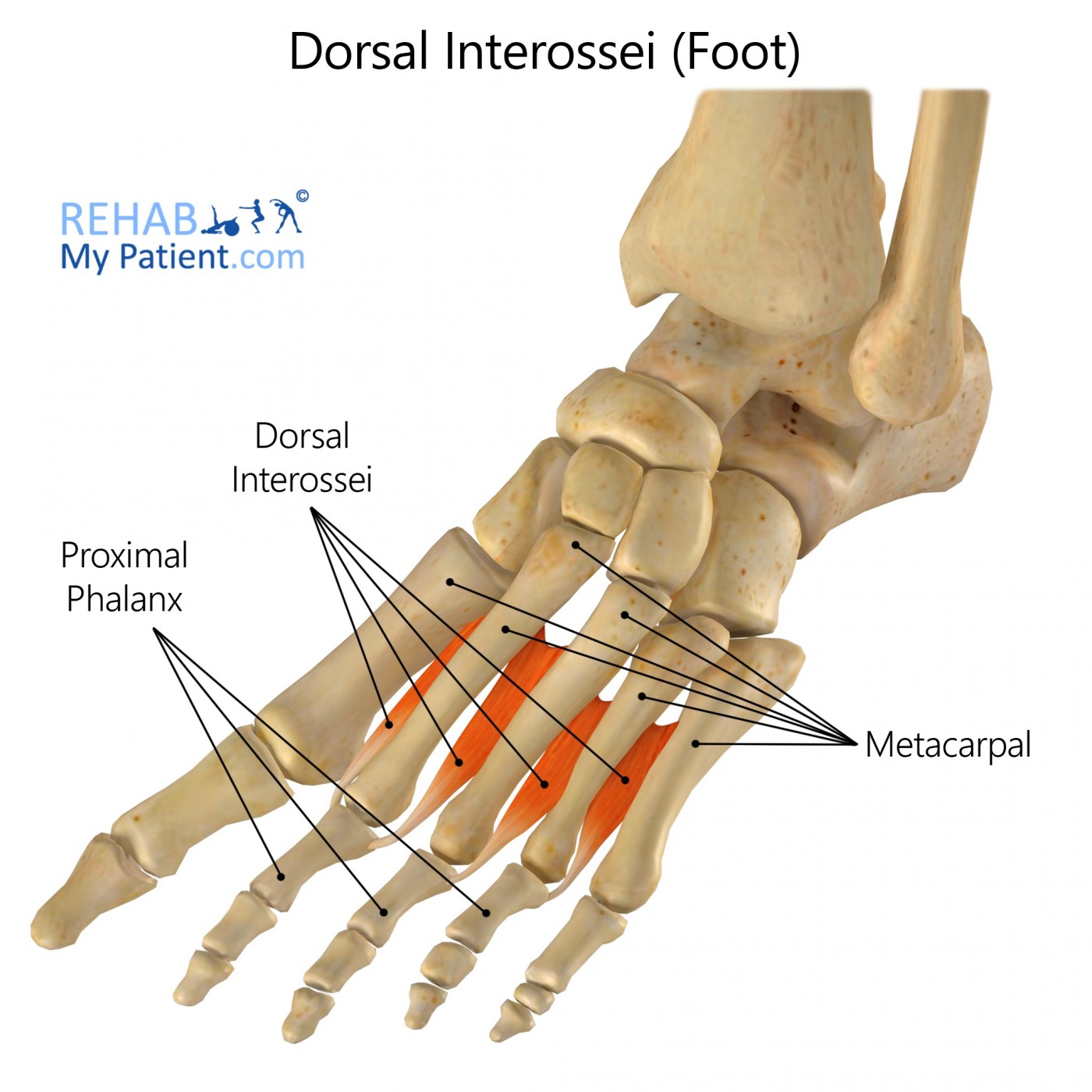
Dorsal Interossei Of The Foot Rehab My Patient from www.rehabmypatient.com In this weeks video, we have a look at muscle edema in the intrinsic and plantar muscles of the foot and what it can mean.patreons can access original dicom. Several rehabilitative exercises have been proposed to improve it; Muscle atrophy in the plantar foot muscles using mri techniques in similar subjects. First & second layers of muscles of the sole 3. Their function lies in spreading the toes apart and in flexing the metatarsophalangeal joints of the second to the fifth toe. To see a 3d model of the dorsal interossei of the foot follow this link. Because there is a pair of dorsal interossei muscles attached on both sides of the second toe, simultaneous contraction of these muscles results in no movement. Muscle mri is useful for the detection of pathological muscles in dm1 patients with gait all dm1 patients presenting with foot drop showed high intensity signals in the tibialis anterior muscles on.
Foot interossei muscles mri / flexor hallucis longus an overview sciencedirect topics / flexors on the medial side.
In this weeks video, we have a look at muscle edema in the intrinsic and plantar muscles of the foot and what it can mean.patreons can access original dicom. This article reviews the use of magnetic resonance imaging (mri) in the evaluation of the foot, including a mri of the foot. The interossei muscles of the foot have two layers, a dorsal layer and a plantar layer. Screen on fatsat images for bone marrow edema. The foot contains many bones, muscles, tendons, and other structures. Anatomy of the whole human body : Their function lies in spreading the toes apart and in flexing the metatarsophalangeal joints of the second to the fifth toe. The extensor digitorum brevis and extensor hallucis brevis arise on the dorsum of the foot. The plantar muscles form three lengthwise groups, which are incompletely separated by connective tissue septa. Characterized by an increase in free water, muscle edema is well depicted by mri. Medial sides of metatarsals of toes iii to v insertion: Originates from the medial and lateral tubercles of the calcaneus and the plantar aponeurosis. Adduction of toes iii to v at metatarsophalangeal joints;
Dorsal interossei (foot) medically reviewed by the healthline medical network — written by the healthline editorial team on january 21, 2018 there are four dorsal interossei muscles in the foot. Since some of the forefoot intrinsic muscles, such as interossei muscles, do not provide direct mechanical support to the medical longitudinal arch, rearfoot muscle strength may therefore be the focus of rehabilitation in the strengthening exercise protocol. As the foot muscles can be divided either from lateral to medial into three groups or from superficial to deep into four layers, plantar interossei can be classified in two ways: Injuries to the metatarsophalangeal joints in athletes. Flexors on the medial side.
Interosseous Muscles Palmar Uw Radiology Muscle Anatomy Hand Therapy Anatomy from i.pinimg.com Attaches to the base of the proximal phalanx of the fifth digit. We use a checklist when evaluating an mri of the ankle: Several rehabilitative exercises have been proposed to improve it; Muscle atrophy in the plantar foot muscles using mri techniques in similar subjects. Coronal images are perpendicular to the long axis of the metatarsals. Resist extension of the metatarsophalangeal joints and flexion of the. It resembles the interossei in structure. They are smaller than the dorsal interossei of the hand.
Anatomy of the whole human body :
We use a checklist when evaluating an mri of the ankle: However, ifm atrophy could be an. Check the syndesmosis, the lateral and medial ligaments. Coronal images are perpendicular to the long axis of the metatarsals. Muscle atrophy in the plantar foot muscles using mri techniques in similar subjects. Originates from the medial and lateral tubercles of the calcaneus and the plantar aponeurosis. The three plantar interossei muscles adduct the 3 rd, 4 th and 5 th toes toward the long axis through the 2 nd toe. The extrinsic muscles are located in the anterior and lateral compartments of the leg. Concerning that the plantar foot muscles can be divided either into layers (superficial to deep) or into groups (medial to lateral), dorsal interossei can be grouped under; They lie between the metatarsals and have some interesting properties. In this weeks video, we have a look at muscle edema in the intrinsic and plantar muscles. Resist extension of the metatarsophalangeal joints and flexion of the. The foot contains many bones, muscles, tendons, and other structures.
The foot contains many bones, muscles, tendons, and other structures foot muscles mri. (there are four layers.) there are four dorsal and 3 plantar muscles.